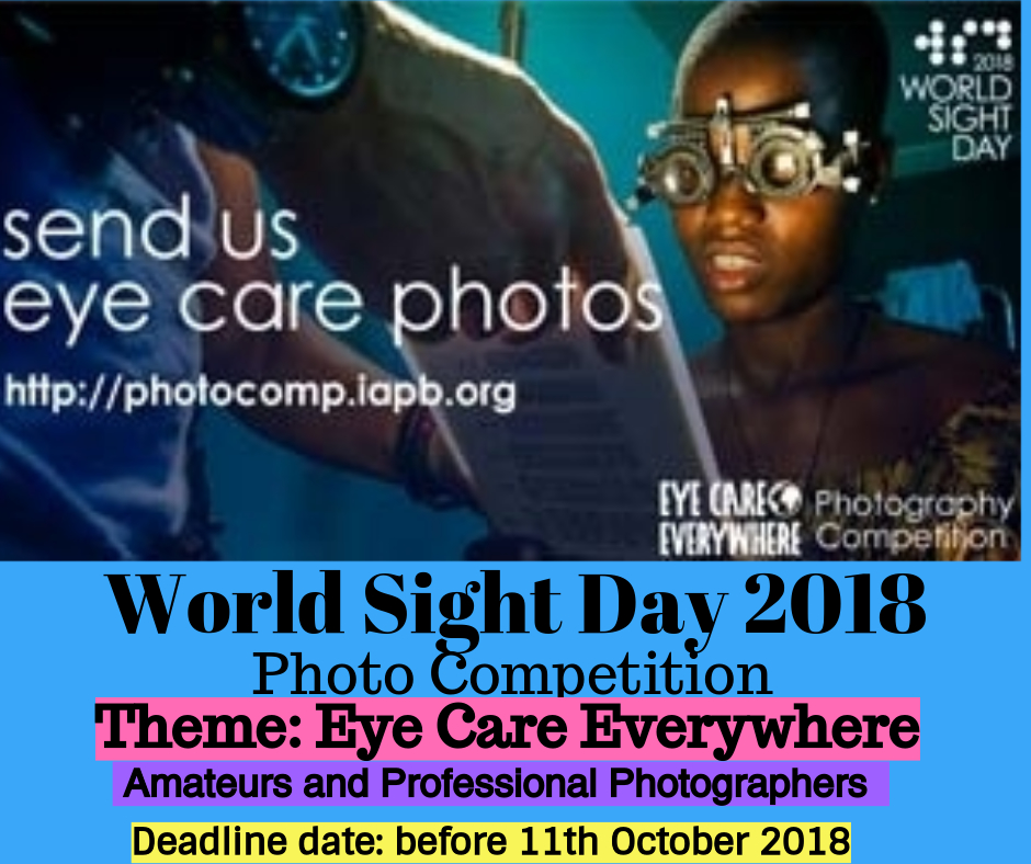
Lets Represent The Caribbean…IAPB Photo Competition
This is a great opportunity for OSWI membership to grab your cameras and let’s represent the Caribbean eye care community. Please note the certain deadlines. There are main prizes and special mention. Do not wait until the deadline. Keep uploading your photos so you can qualify for the special mention prizes which end on 15th -16th September 2018. Competition ends on 11th October 2018
After the immense success of their three successive photo competitions, the International Agency for the Prevention of Blindness (IAPB) invites amateur and professional photographers from around the world to join them in celebrating the diversity of stakeholders and patients they reach. With support from Bayer Health Care, the theme for the international Photography Competition this year is ‘Eye Care Everywhere’.

“Eye health work across the world is inspiring change and making it a better place”, notes Joanna Conlon, Director of Development and Communications. “These pictures shine a light on the impact eye care organisations, including IAPB members, have around the world. Together, we can ensure that everyone everywhere experience positive change.”
Theme
1.2 billion people don’t have access to glasses. Over 3 out of 4–more than 75%–of the world’s vision impaired are avoidably so. The photo competition is a great tool to draw a spotlight on eye care issues across the globe and those who are vulnerable to sight conditions—young children, persons with diabetes, older people, people with limited access to health services etc. With the theme, “#EyeCareEverywhere”, IAPB encourages you to share images that highlight the impact, and strength of our partnerships and most importantly highlight the importance of vision everywhere in the world.
World Sight Day (11 October 2018) is an international day of awareness about avoidable blindness and its prevention and is an important advocacy and communications opportunity for the eye health community. It is a great time to engage with a wider audience – a patient’s family, those who seldom get an eye exam, diabetics – and showcase why eye health needs everybody’s attention.
Participation in the competition is open worldwide – upload a photo that best exemplifies eyecare everywhere, give it a title or caption; clearly note your name, profession and contact details on the competition micro-site: http://photocomp.iapb.org. You can read the competition guidelines, here.
Prizes
All individuals interested in the theme are welcome to participate in the photo competition. Send us your photos with the theme ‘Eye Care Everywhere’ before World Sight Day (11th October 2018). Every week, IAPB will pick “Editor’s picks” and showcase them on IAPB’s social media accounts. Finally, winners will be selected from these Editor’s picks after World Sight Day (the last day of the competition). The competition will be open till 11 October 2018 – World Sight Day, after which IAPB will pick two winners and 5 runners-up. Winners will be announced on 15 October 2018. The Amateur prize will be a professional camera: Canon 1200 D DSLR camera. The professional prize this year will be a cash prize of USD 1000.
Special Prize
This year, we have a special prize: delegates to the IAPB Council of members in Hyderabad, India will vote for their most favoured submission from the Editor’s picks till the Council begins. The dates of the Council are 15-16 September 2018. The winner will get a special mention and a certificate.
For more information do visit the micro-site: http://photocomp.iapb.org
READ MORE
WIGLS Takeaway 24 Months Data
[et_pb_section admin_label=”section”][et_pb_row admin_label=”row”][et_pb_column type=”4_4″][et_pb_text admin_label=”Text” background_layout=”light” text_orientation=”left” use_border_color=”off” border_color=”#ffffff” border_style=”solid”]
Take-away from WIGLS 24 month data:
1. SLT safely and effectively lowers IOP in Afro-Caribbeans with POAG.
2. Most patients will not need medications.
3. SLT can be safely repeated when its initial effect wanes.
4. SLT should be considered a preferred primary therapy for POAG in people of African descent.
Tony Realini
Take away from Transcleral CPC in eyes with good vision:
1. Viable, safe, easy primary procedure for eyes with uncontrolled IOP
2. Loss of vision associated with higher energy settings
3. Head to head comparison between micropulse cpc and continuous wave cpc also cpc vs
conventional glaucoma filtration surgery needed.
Arindel Maharaj MD, PhD
EBAA High Impact Research Grant
[et_pb_section admin_label=”section”][et_pb_row admin_label=”row”][et_pb_column type=”4_4″][et_pb_text admin_label=”Text” background_layout=”light” text_orientation=”left” use_border_color=”off” border_color=”#ffffff” border_style=”solid”][/et_pb_text][/et_pb_column][/et_pb_row][/et_pb_section] READ MORENew Grant Opportunity!EBAA High Impact Research GrantDeadline: Friday, September 14EBAA is currently accepting proposals for the 2018 EBAA High Impact Research Grant. The EBAA High Impact Research Grant is a new grant opportunity offered by EBAA in an effort to make a significant impact on cornea and eye banking research. Each year, EBAA will fund a high impact project focused on a specific topic of interest to the eye banking and corneal transplant community. This year, EBAA is requesting proposals on “Maintaining Endothelial Viability During Tissue Processing and Transport.” One proposal will be selected for funding in 2018 up to $50,000. Applications are due September 14. For the opportunity to be selected for this exciting new grant; submit a proposal today!If you have any questions about either opportunity, please contact Stacey Gardner with any questions.
Cornea and Eye Banking Forum
[et_pb_section admin_label=”section”][et_pb_row admin_label=”row”][et_pb_column type=”4_4″][et_pb_text admin_label=”Text” background_layout=”light” text_orientation=”left” use_border_color=”off” border_color=”#ffffff” border_style=”solid”]Abstracts Due Monday!
Cornea and Eye Banking Forum
Friday, October 26, 2018
Chicago, IL
Abstract Submissions Due August 6
Early Bird Registration Ends September 7
Abstract submission for the Cornea and Eye Banking Forum, jointly hosted by EBAA and Cornea Society, closes on Monday, August 6. The forum will take place Friday, October 26 at the Westin Michigan Avenue, and will showcase the presentation of at least 25 scientific papers. This annual forum has long been favored by ophthalmologists and eye bankers as it continues to feature the latest research and information in the fields of corneal transplantation and eye banking.
Here is what last year’s attendees said about the meeting’s educational content:
- “Great papers presented. Only meeting where I leave and change something about how I practice medicine.”
- “Best true cornea scientific meeting of the year!”
- “I think it has the most cutting-edge research, and comments from thought leaders in cornea are invaluable.”
- “Innovative and topical presentations related to cornea transplant and eye banking.”
- “The information shared here is way ahead of anything published.”
If you have conducted research in cornea transplantation, disease, preservation, or preparation and are interested in presenting to the leaders in the fields of cornea transplantation and eye banking, we encourage you or your Fellows and Residents to share research results and clinical findings by submitting an abstract for inclusion in the program. All corneal residents and fellows who present their abstracts will be eligible for the Best Paper of Session Award, that includes a monetary award of $1,500. The deadline to submit an abstract is Monday, August 6, 2018. For more information or to register for the Forum, click here.
[/et_pb_text][/et_pb_column][/et_pb_row][/et_pb_section] READ MOREMIGS take away points
[et_pb_section admin_label=”section”][et_pb_row admin_label=”row”][et_pb_column type=”4_4″][et_pb_text admin_label=”Text” background_layout=”light” text_orientation=”left” use_border_color=”off” border_color=”#ffffff” border_style=”solid”]
It’s not just trabs and tubes anymore: from most invasive to microinvasive
TAKE AWAY POINT No. 1: Poor adherence is common
Poor adherence to treatment is relatively common among glaucoma patients and is associated with progression of disease. Quite often, medical treatment for glaucoma is suboptimal.
TAKE AWAY POINT No. 2: There are four routes by which to surgically lower IOP
1. increase trabecular outflow
2. increase uveoscleral outflow
3. decrease aqueous fluid production
4. increase aqueous outflow into the subconjunctival space
TAKE AWAY POINT No.3: The term MIGS was coined in 2009 by Dr. Ike Ahmed
There is currently no single common and widely accepted definition of MIGS. It was originally known as “minimally invasive” but has since evolved to “microinvasive”.
According to Saheb and Ahmed, the term MIGS refers to a group of surgical procedures which share five preferable qualities (Saheb H, Ahmed II. Micro-invasive glaucoma surgery: current perspectives and future directions. Curr Opin Ophthalmol. 2012;23(2):96–104):
1. an ab interno approach through a clear corneal incision which spares the conjunctiva of incision
2. a minimally traumatic procedure to the target tissue
3. an IOP lowering efficacy that justifies the approach
4. a high safety profile avoiding serious complications compared to other glaucoma surgeries
5. a rapid recovery with minimal impact on the patient’s quality of life.
TAKE AWAY POINT No. 4: MIGS is NOT a replacement for filtration surgery.
Filtration surgery is still indicated in:
1. angle closure glaucoma
2. when very low target IOP are needed: severe/advanced glaucoma and normal tension
glaucoma
TAKE AWAY POINT No. 5: Treatment algorithm
Mild glaucoma – Pharmacotherapy and selective laser trabeculoplasty (SLT)
Moderate glaucoma – MIGS (combined with cataract extraction or stand alone)
Severe glaucoma – Filtration surgery +/- cataract extraction (trabeculectomy +/- anti-metabolite and tube shunt surgery)
[/et_pb_text][/et_pb_column][/et_pb_row][/et_pb_section] READ MOREGrading of Angle Width
[et_pb_section admin_label=”section”][et_pb_row admin_label=”row”][et_pb_column type=”4_4″][et_pb_text admin_label=”Text” background_layout=”light” text_orientation=”left” use_border_color=”off” border_color=”#ffffff” border_style=”solid”]Grading of Angle Width
The grading of angle width is an essential part of the ocular examination.
The main aims are to evaluate the functional status of the angle, the
degree of closure, and the risk of further closure. It is important to
determine:
1. the geometrical angle width in degrees
2. the shape and contour of the peripheral iris
3. the most posterior structure seen
4. the presence of peripheral anterior synechiae
5. the amount of trabecular pigmentation.
Shaffer Grading System
The Shaffer system (Fig. 13.13) records the angle in degrees of arc
subtended by two imaginary tangential lines drawn to the inner surface
of the trabecula and the anterior surface of the iris about one-third of the
distance from its periphery. 10 In practice, the angle is graded
according to the visibility of various angle structures. The
system assigns a numerical grade (4–0) to each angle with associated
anatomical description, angle width in degrees, and implied clinical
interpretation.
1. Grade 4 (35–45°) is the widest angle characteristic of myopia and
aphakia in which the ciliary body can be visualized with ease; it is
incapable of closure.
2. Grade 3 (25–35°) is an open angle in which at least the scleral spur can
be identified; it is also incapable of closure.
3. Grade 2 (20°) is a moderately narrow angle in which only the trabecula
can be identified; angle closure is possible but unlikely.
4. Grade 1 (10°) is a very narrow angle in which only Schwalbe’s line, and
perhaps also the top of the trabecula, can be identified; angle closure is
not inevitable but the risk is high.
5. Slit angle is one in which there is no obvious iridocorneal contact but no
angle structures can be identified; this angle has the greatest danger of
imminent closure.
6. Grade 0 (0°) is a closed angle due to iridocorneal contact and is
recognized by the inability to identify the apex of the corneal wedge.
Indentation gonioscopy with a Zeiss goniolens is necessary to
differentiate ‘appositional’ from ‘synechial’ angle closure.
FIGURE13.13
Shaffer grading system (see text).
Reproduced from Salmon JF, Kanski JJ, eds

In real life the degree measurement of the angle eg 20 degrees, 30 degrees, 40 degrees might not always exactly correspond to the structures seen. For example, you might be able to see as far as scleral spur, thus designating the angle grade 3, but the actual “walls” of the angle (if you think of it like a valley between the cornea and iris or TM tangent and iris) might be quite steep and deep, so it could be 20 degrees rather than the 30 degrees you would expect with a grade
3. This scenario is uncommon, and usually the angle between the “valley walls” corresponds to the structures seen, but of course, patients don’t read the textbook. The important message about angle grading with Shaffer is to grade according to the structures seen, from Grades 0 to
4. You will notice in the attached Word excerpt from Glaucoma by Shaaraway et al., that the degree measurement is in brackets as a secondary notation. Important when recording to note not just the Shaffer number, but also the structures seen, the degree measurement, and the
presence or absence of other features such as PAS, pigment, vessels, masses.
